About the Images
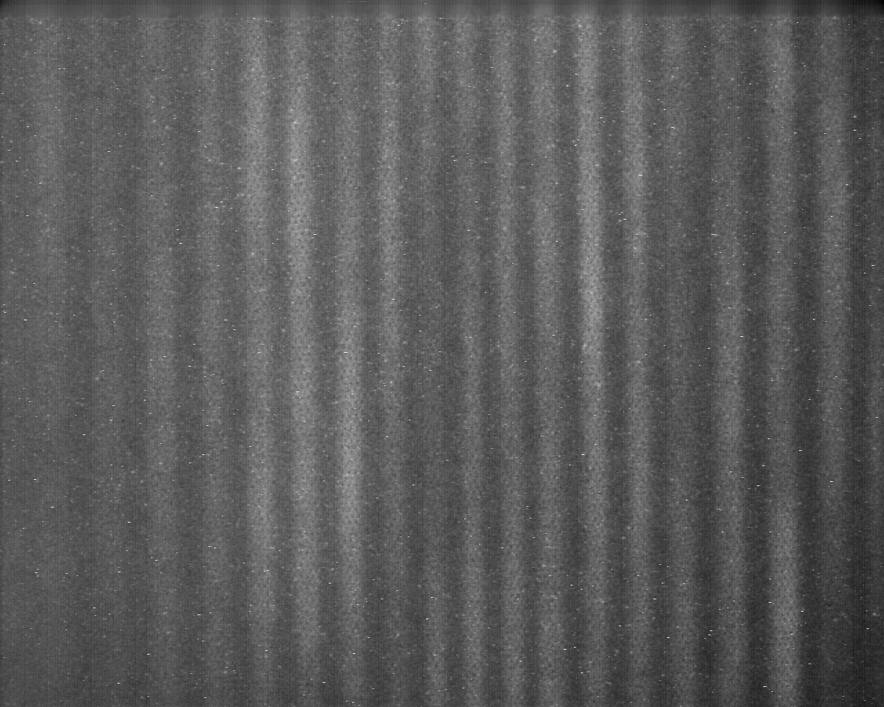
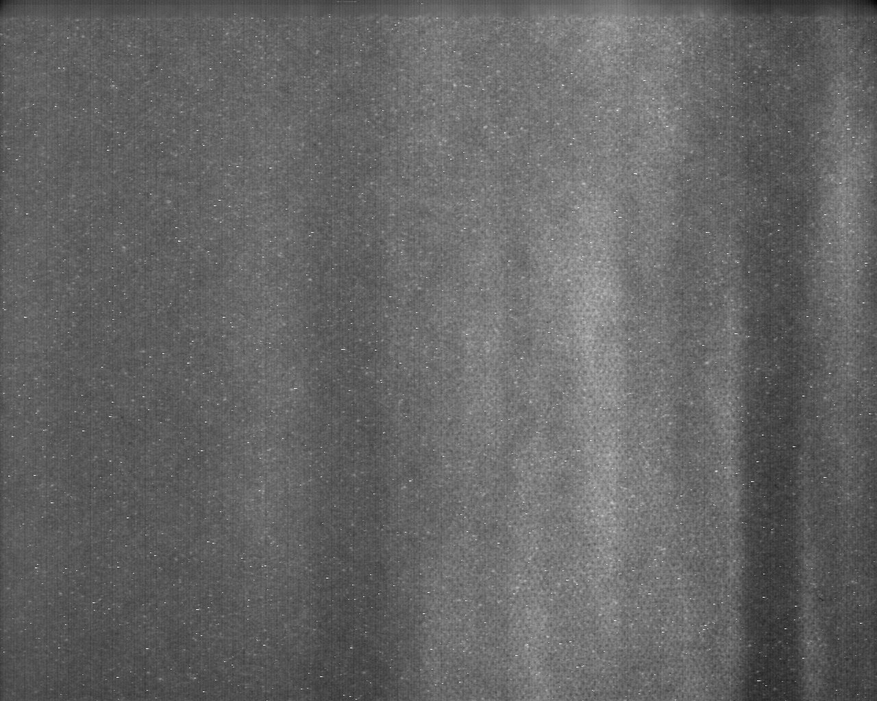
These upper two images show an x-ray camera image of a graphite reflector, with and without an opaque pattern overlay. These are thought to be the first detailing the inhomogeneous nature of crystalite alignment/spacing in H.O.P.G. - the most common monochromator used in powder diffraction. This data indicates a need for sample placement optimization for non-uniform materials.
The field-of-view of the compound refractive x-ray lens (as viewing the reflector) is 2.5-mm. Illuminating radiation is ~17.5 keV. Reflector to lens distance is 850-mm. Lens to detector distance is 1.28-m. Magnification is 1.5X. Detector resolution is 10-microns.
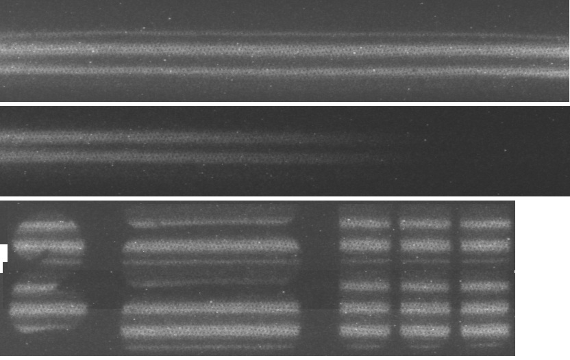
Composite image of test pattern grids: top, 50-micron bar magnified 1.6X, middle, edge of multilayer reflection illumination field, bottom, overlay of near-detector pattern as check on image distortion.
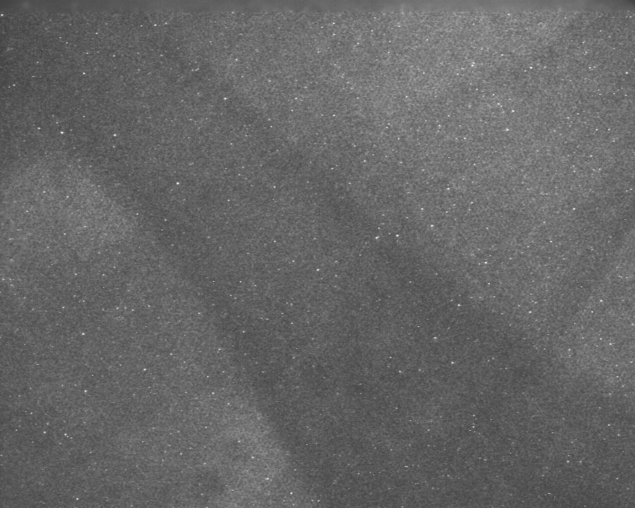
Lastly, a monochromatic radiograph of murine vertebral disc (ex-vivo, whole body image).